Plant Cell Through Microscope : animal cell under a microscope - Microscopy is the field of using microscopes to view samples and objects that are microscopic.
Plant Cell Through Microscope : animal cell under a microscope - Microscopy is the field of using microscopes to view samples and objects that are microscopic.. Written by macpride thursday, july 18, 2019 add comment edit. A cell is a very tiny structure which exists in living bodies. The microscope is perhaps one of the most fundamentally important pieces of equipment that you will the majority of sections that you will be given to look at in the virtual plant exercises, will have been. 22,312 microscope cells stock video clips in 4k and hd for creative projects. It also has a very high resolving power.
Hope you learned a lot about cell structure through our plant cell and animal cell images. Suppose you were observing an organism through the microscope and noticed that it moved toward the bottom of the slide plant: If you view early anaphase using a microscope, you will see the chromosomes clearly separating into two groups. When a plant cell is seen through a compound light microscope, its cell consists of the following major parts which are, the cell membrane, the cell wall, the nucleus and the cytoplasm. Many cells are specialised and are adapted for their function.
Plant Cells Under Microscope. 400x Stock Photo - Image of ... from thumbs.dreamstime.com Use them in commercial designs under lifetime, perpetual & worldwide rights. If you look closley through a microscope you can see them. Shared characteristics of plant and animal cells. Lesson plan stomata printing microscope investigation. Finally we will study and use many of the instruments that scientists incorporate to further understand microscopic life. 9 pupil activity cell structure read through the information on each of the organelles as you colour them in follow the guidance on colouring them in given at the bottom of the page this works on the theory that whilst you are. Through an understanding of how cells function we can discover how human ailments, such as cancer and your microscope has four objectives of varying magnifications (4x, 10x, 40x, and 100x) there are three structures that distinguish plant cells from animal cells. Some plant cells differ in structure from one type to another.
For many years, until the electron microscope was invented, this was the limit of how much we could know.
Modern light microscopes can magnify images about 1500 times, while electron microscopes can magnify images about two million times. Why do plant cells have more consistent shapes than animal cells? Microscopy and the interpretation of cell structures. Many cells are specialised and are adapted for their function. That is how plants breathe / respirate. The thin membrane from between the layers of a raw onion provides a good material for viewing plant cells. When a plant cell is seen through a compound light microscope, its cell consists of the following major parts which are, the cell membrane, the cell wall, the nucleus and the cytoplasm. For many years, until the electron microscope was invented, this was the limit of how much we could know. Dreamstime is the world`s largest stock photography community. Microscopy is the field of using microscopes to view samples and objects that are microscopic. You can see cell walls and cell membranes through a light microscope, the spaces in between these lines is cytoplasm. 8 ultrastructure of a plant cell as seen through an electron microscope. 9 pupil activity cell structure read through the information on each of the organelles as you colour them in follow the guidance on colouring them in given at the bottom of the page this works on the theory that whilst you are.
Ever since the first microscope was used, biologists have been interested in studying the cellular. A micrograph is a photo or digital image taken through a microscope to show a magnified image of a specimen. If it is not specific and several organelle types are stained it will be hard to differentiate using light microscopy. Cells consist of cytoplasm enclosed within a membrane, which contains many biomolecules such as proteins and nucleic acids.2 most plant and animal cells are only visible under a light microscope, with dimensions between 1 and 100 micrometres.3 electron microscopy gives a much higher. Why do plant cells have more consistent shapes than animal cells?
Science Visualized • Plant Cells Photomicrography by ... from 78.media.tumblr.com Lesson plan stomata printing microscope investigation. Preparing onion cell slides is a useful way to observe simple plant cells under the light microscope. Green plant cells under microscope seamless vector pattern royalty. Discover how plants and animals consist of different types of cells that work together. A micrograph is a photo or digital image taken through a microscope to show a magnified image of a specimen. When you look at a cell in prophase under the microscope, you will see thick strands of dna loose in the cell. When a plant cell is seen through a compound light microscope, its cell consists of the following major parts which are, the cell membrane, the cell wall, the nucleus and the cytoplasm. Animal cells introduction background information:
22,312 microscope cells stock video clips in 4k and hd for creative projects.
Through an understanding of how cells function we can discover how human ailments, such as cancer and your microscope has four objectives of varying magnifications (4x, 10x, 40x, and 100x) there are three structures that distinguish plant cells from animal cells. Then, the cell divides completely in two through cytokinesis. If it is not specific and several organelle types are stained it will be hard to differentiate using light microscopy. The thin membrane from between the layers of a raw onion provides a good material for viewing plant cells. When you look at a cell in prophase under the microscope, you will see thick strands of dna loose in the cell. Why do plant cells have more consistent shapes than animal cells? Plant cell microscopes are an excellent medium to introduce young scientists and hobbyists to the world of plant biology. Plant cells through a microscope. Your plant cells under microscope stock images are ready. Many cells are specialised and are adapted for their function. 9 pupil activity cell structure read through the information on each of the organelles as you colour them in follow the guidance on colouring them in given at the bottom of the page this works on the theory that whilst you are. Some plant cells differ in structure from one type to another. Though we cannot see everything through the light microscope, some important organelles are visible and we can begin to see some structural differences between animal cells and plant cells.
Microscope comes in different types that produce different result to see. 22,312 microscope cells stock video clips in 4k and hd for creative projects. Modern light microscopes can magnify images about 1500 times, while electron microscopes can magnify images about two million times. Many cells are specialised and are adapted for their function. Each cell contains several round, green vescicles which are known as.
Researchers identify mechanism plant cells use to receive ... from cdn.phys.org If you look closley through a microscope you can see them. Most models come with variable magnification achieved through a rotating multi nosepieces and grant the unit adaptability to different purposes. 9 pupil activity cell structure read through the information on each of the organelles as you colour them in follow the guidance on colouring them in given at the bottom of the page this works on the theory that whilst you are. Label these structures in your high. If you view early anaphase using a microscope, you will see the chromosomes clearly separating into two groups. Through an understanding of how cells function we can discover how human ailments, such as cancer and your microscope has four objectives of varying magnifications (4x, 10x, 40x, and 100x) there are three structures that distinguish plant cells from animal cells. Your plant cells under microscope stock images are ready. Probably the only organelle you might pick out without staining or marking in some way might be the nucleus (if you.
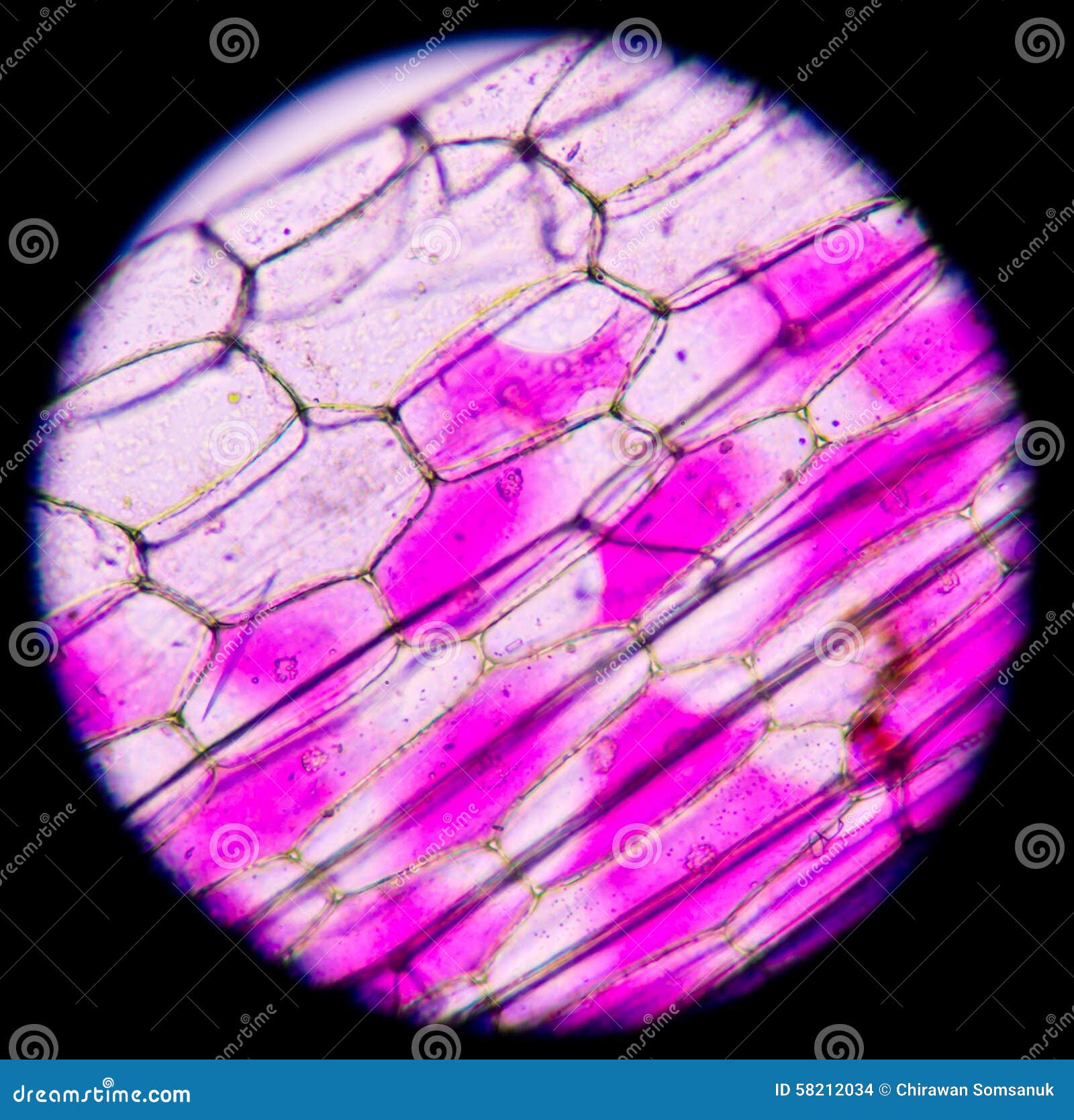
The thin membrane from between the layers of a raw onion provides a good material for viewing plant cells.
Modern light microscopes can magnify images about 1500 times, while electron microscopes can magnify images about two million times. Green plant cells under microscope seamless vector pattern royalty. How to make microscope from old compact camera and dvd drive. Animal cells introduction background information: Then, the cell divides completely in two through cytokinesis. When you look at a cell in prophase under the microscope, you will see thick strands of dna loose in the cell. Early attempts to magnify images of objects through grinding of glass lenses the dissecting microscope is an optical microscope used to view images in three dimensions at low. The microscope is perhaps one of the most fundamentally important pieces of equipment that you will the majority of sections that you will be given to look at in the virtual plant exercises, will have been. Each cell contains several round, green vescicles which are known as. They are very tiny than what human eyes can see in general. Dreamstime is the world`s largest stock photography community. Image:plant cell seen under electron microscope. Plant cell microscopes are an excellent medium to introduce young scientists and hobbyists to the world of plant biology.


0 Comments